Martha Marciano /CONECTA
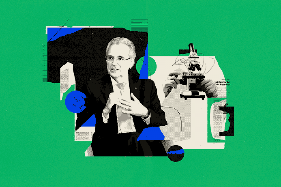
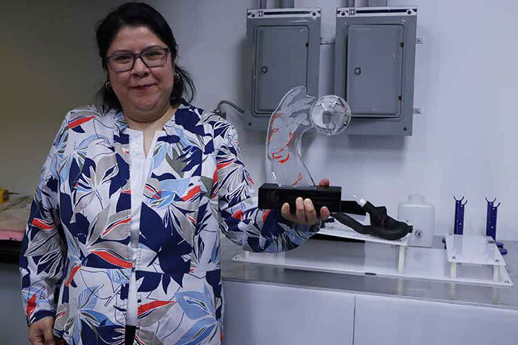
Researcher Erika García-López from the School of Engineering and Sciences has received the 2024 Tecnos Nuevo León 4.0 Award for developing a cardiac simulator which accelerates the recovery of patients suffering from heart disease.
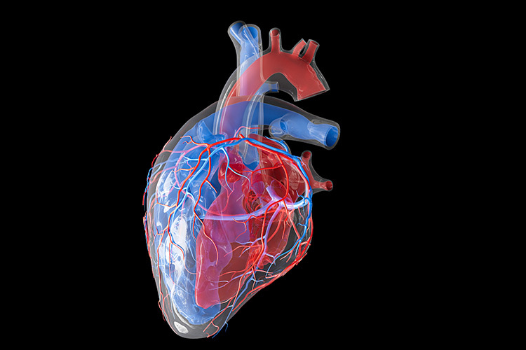
“As the most common cause of death around the world continues to be heart disease, we started working on a simulator. TAVI (Transcatheter Aortic Valve Implantation) consists of placing an implant in the aorta of patients, particularly those over 60 years old,” García-López explained.
“Heart Catgh Simulator”, which is an auxiliary medical device for cardiac procedures, is the name of the Tec de Monterrey project that won a prize in the “Human Coexistence in an Inclusive City” category.
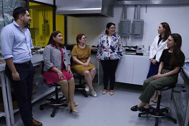
The research group led by García-López includes students and specialists in Mechatronics, Industrial, Electronics, Mechanics, and other fields found on the Monterrey campus.
“This project brings us to the forefront of science; we competed in the Tecnos Award because the project incorporates elements of digitization, manufacturing, sensors, and even cloud-based data acquisition,” shared the teacher.
How Does the Device Work?
The medical procedure known as TAVI consists of inserting the aortic valve through catheters, meaning that the sternum does not have to be opened (sternotomy), thus accelerating patients’ recovery.
“In the aorta, there is a type of valve cusp which calcifies due to age and causes the valve to no longer have the same opening and closing ability, so a device is inserted,” García-López explained.
The valve implant comprises 3 parts: a mesh that serves as a structure, three valve cusps that open and close, and a cover that surrounds the mesh.
Initially, the team planned to undertake catheter navigability tests before inserting the implant. The studies were carried out in collaboration with the Imaging Department at TecSalud’s Zambrano Hellion Hospital and made use of real tomography scans.
This team digitized the tomography scans using different software to adapt them into a 3D-printed anatomical model.
“Although we’d already been working on the simulator within the research group, there came a point in the development when we needed to do more conceptual testing of how the expansion was going,” she added.
Simulator Could Help Paramedics and Students
The device is in the testing stage and is currently being used on different segments of patients; the work on digitizing new anatomical models continues.
“Thanks to the support of the institution, we incorporated a flow pump into our anatomical model, so we now have a complete and functional cardio system. It includes femoral arteries that reach the aorta, and we’ve even been able to create a heartbeat”, said the professor.
The researcher aims to create products that serve the community and believes that the simulator project will positively impact and serve paramedics and medical students who are training.
